:::

A powerful tool for X-ray imaging technology development – the multi-functional X-ray imaging platform


Published:2025-07-23  15:25

【Technical Introduction】
X-ray imaging is widely used in medical diagnostics and industrial non-destructive testing (NDT) because X-ray photons can penetrate objects to reveal internal structures without physical sectioning or damage. In Taiwan, however, most X-ray imaging equipment is imported, and key specifications and operating parameters are constrained by original manufacturer designs, limiting flexibility for diverse inspection requirements.
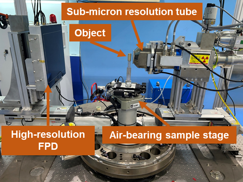
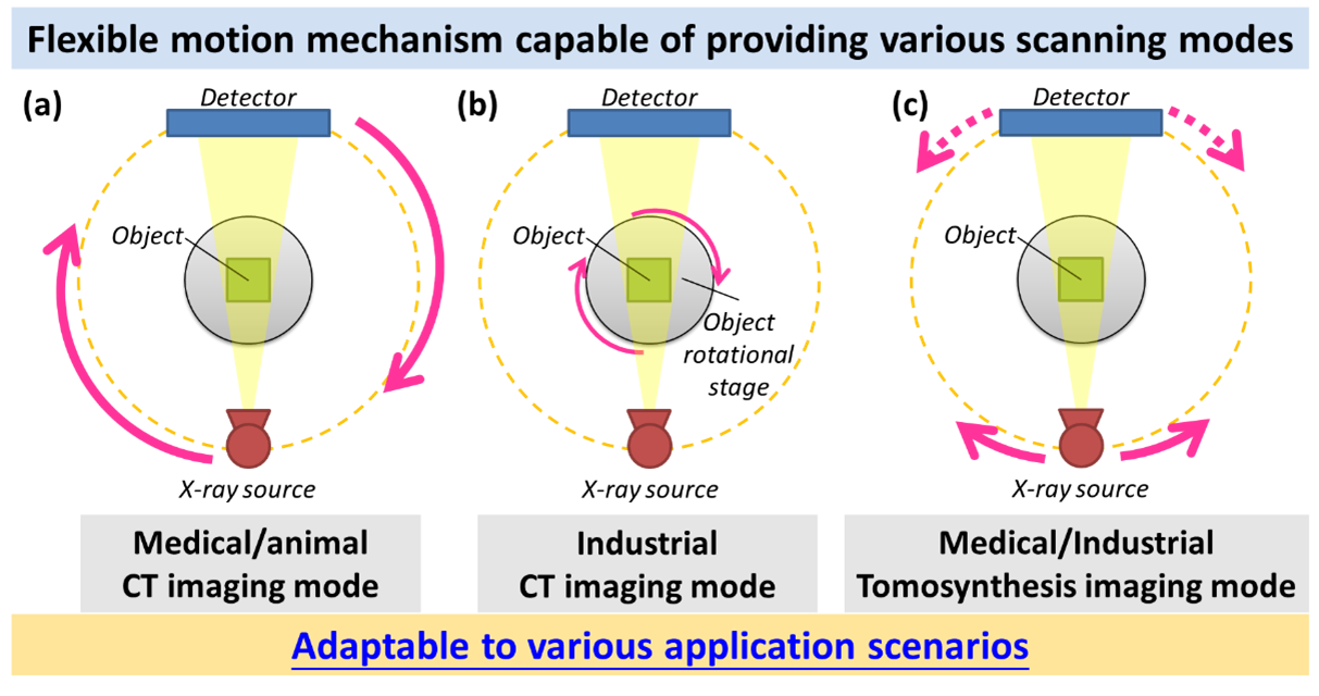
To address this gap, NARI developed a multifunctional X-ray imaging platform equipped with a 7-axis motion system to enable multiple scanning modes and rapid reconfiguration through interchangeable key components. The platform further integrates proprietary data acquisition, image calibration, and 3D reconstruction algorithms, providing a robust environment for proof-of-concept demonstrations, system parameter optimization, and efficient development of new X-ray systems and components—reducing both development time and cost.

Figure 1. Photograph of the NARI-developed multifunctional X-ray imaging platform.


Figure 2. Schematic of the multi-functional X-ray imaging platform's flexible motion mechanism capable of various scanning modes.

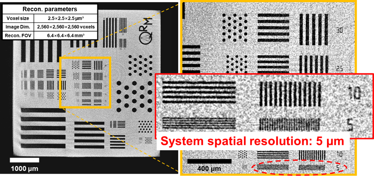
Figure 3. Micro-CT spatial resolution verification. Reconstructed images of a QRM test pattern demonstrate resolvable features down to 5 μm with a 2.5 μm isotropic voxel size, indicating an approximately 5 μm system spatial resolution.

Sandstone pore network revealed by NARI micro-CT. Reconstructed cross-sections and magnified views (A, B) clearly visualize internal pore structures in an approximately 5 mm sandstone specimen.

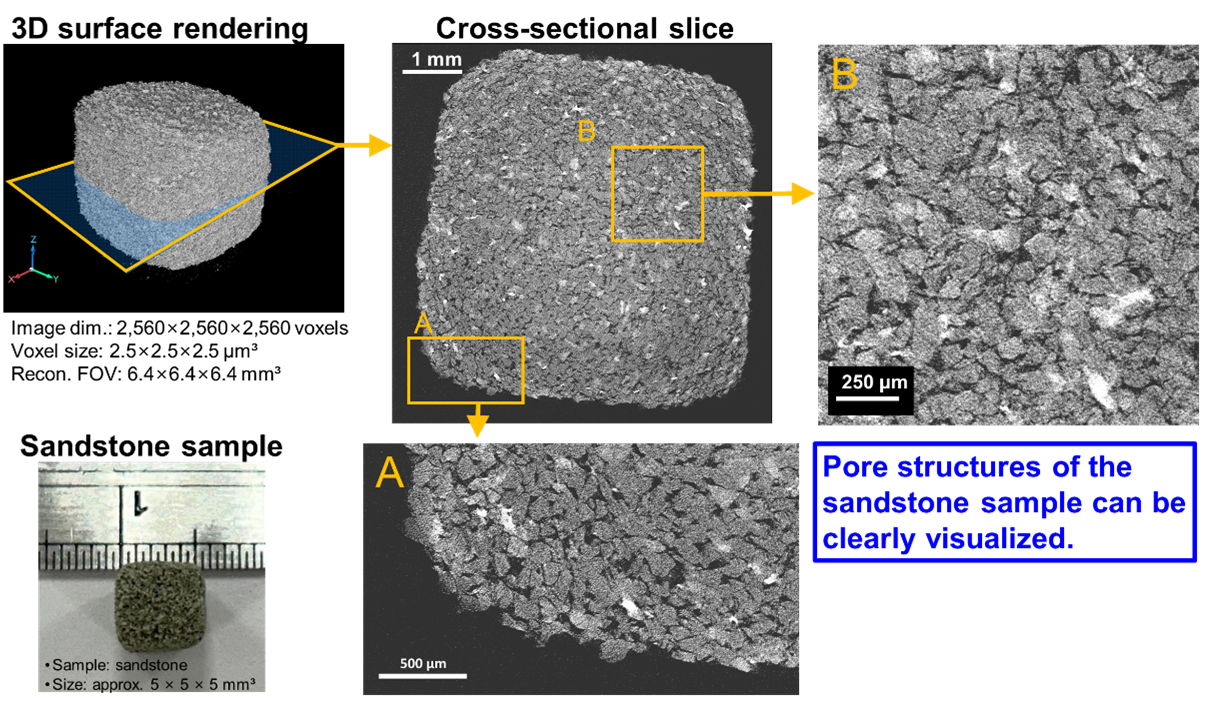
Figure 4. Sandstone pore network revealed by NARI micro-CT. Reconstructed cross-sections and magnified views (A, B) clearly visualize internal pore structures in an approximately 5 mm sandstone specimen.

【Project Planning/Technical Application】
The multi-functionality of this platform enables itself to support various X-ray imaging scanning modes, making it be applicable for across a wide range of medical and industrial fields. These applications include medical chest imaging, orthopedics, dentistry, mammography, veterinary scans, as well as industrial non-destructive testing (NDT) and security inspections. The platform is highly effective in assisting those organizations engaged in the development of X-ray imaging technologies by facilitating concept validation and system parameter optimization. This capability significantly shortens the product development cycles and reduces associated costs. Furthermore, the platform offers measurement services for critical X-ray imaging performance indicators of the key components, such as modulation transfer function (MTF) and noise power spectrum (NPS).
 
【Future layout】
Building on the foundation of this platform, NARI will continue advancing the development of the cutting-edge X-ray imaging technologies, including spectral radiography and material decomposition, high-resolution 3D-CT, and high-energy X-ray imaging. Moving forward, NARI plans to collaborate with domestic academic, research, and medical institutions to further broaden the applications of X-ray imaging technology. The goal is to enhance the competitiveness of domestic X-ray imaging technologies and contribute to the growth of a robust, X-ray-related industry.
 
【Contact information】
Name: Chia-Hao Chang
Tel: 03-4711400 extension 7763
E-mail: chhchang@nari.org.tw